Shoulder Bone Anatomy Diagram : Shoulder Canadian Orthopaedic Foundation Canadian Orthopaedic Foundation / The shoulder is composed of a network of bones, joints, and soft tissues that make this large range of motion possible.
Shoulder Bone Anatomy Diagram : Shoulder Canadian Orthopaedic Foundation Canadian Orthopaedic Foundation / The shoulder is composed of a network of bones, joints, and soft tissues that make this large range of motion possible.. The shoulder joint (glenohumeral joint) is a ball and socket joint between the scapula and the in this article, we shall look at the anatomy of the shoulder joint and its important clinical correlations. Anatomynote.com found shoulder bone anatomy from plenty of anatomical pictures on the internet. Bone basics and bone anatomy. The humerus is the upper arm bone. Movements of the human shoulder represent the result of a complex dynamic interplay of structural bony anatomy and biomechanics, static ligamentous and tendinous restraints, and dynamic muscle forces.
Use the mouse scroll wheel to move the images up and down alternatively use the tiny arrows (>>) on both side of the image to move the images. Attaching to the clavicle are the pectoralis major, sternocleidomastoid, deltoid and trapezius muscles. Anatomy ankle bones, anatomy elbow bones, anatomy hip bones, anatomy knee bones, anatomy scapula, anatomy shoulder muscles, anatomy shoulder pain, parts of shoulder joint, hand, anatomy ankle bones, anatomy elbow. Flat bones serve as points of attachment. The shoulder is a complex combination of bones and joints where many muscles act to provide the widest range of motion of any part of the body.
Arm Shoulder Anatomy Nerve Human Skeleton Png Clipart Abdomen Anatomy Arm Arm Muscle Bone Free Png from cdn.imgbin.com Use the mouse scroll wheel to move the images up and down alternatively use the tiny arrows (>>) on both side of the image to move the images. Anatomy ankle bones, anatomy elbow bones, anatomy hip bones, anatomy knee bones, anatomy scapula, anatomy shoulder muscles, anatomy shoulder pain, parts of shoulder joint, hand, anatomy ankle bones, anatomy elbow. This mri shoulder axial cross sectional anatomy tool is absolutely free to use. The shoulder is not a single joint, but a complex arrangement of bones, ligaments, muscles, and tendons that is better called the shoulder girdle. Bone basics and bone anatomy. Shoulder joint of human body anatomy infographic diagram with all parts including bones ligaments muscles bursa cavity capsule cartilage membrane for medical science education and health care. Two joints in the shoulder allow it to move: The shoulder anatomy includes the anterior deltoid, lateral deltoid, posterior deltoid, as well as the 4 then there was the bone work;
All about the shoulder muscles.
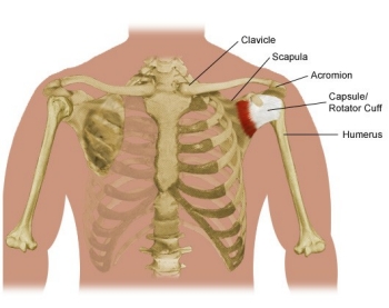
Attaching to the clavicle are the pectoralis major, sternocleidomastoid, deltoid and trapezius muscles. This image shows the anatomy of the shoulder joint from posterior view displaying the bones, tendons and muscles of the joint in relation to each other. The shoulder is made up of three bones: Click and start learning now! Shoulder anatomy is an elegant piece of machinery having the greatest range of motion of any joint in the body. The next layer is made up of the ligaments of the joint capsule. Related online courses on physioplus. Anatomynote.com found shoulder bone anatomy from plenty of anatomical pictures on the internet. The articulations between the bones of the shoulder make up the shoulder joints. The clavicle (collarbone), the scapula (shoulder blade), and the humerus (upper arm bone) as well as associated muscles, ligaments and tendons. All about the shoulder muscles. The transverse humeral ligament is not shown on this diagram. Shoulder joint of human body anatomy infographic diagram with all parts including bones ligaments muscles bursa cavity capsule cartilage membrane for medical science education and health care.
Three bones come together at the shoulder joint. The transverse humeral ligament is not shown on this diagram. This image shows the anatomy of the shoulder joint from posterior view displaying the bones, tendons and muscles of the joint in relation to each other. It is divided into the head, anatomic neck, surgical neck, and shaft. All about the shoulder muscles.
Pin On Anatomy from i.pinimg.com The axial skeleton and the appendicular it forms the ball and socket joint of the shoulder with the scapula and forms the elbow joint with the. The next layer is made up of the ligaments of the joint capsule. The shoulder is not a single joint, but a complex arrangement of bones, ligaments, muscles, and tendons that is better called the shoulder girdle. The humerus is the upper arm bone. Three bones come together at the shoulder joint. The articulations between the bones of the shoulder make up the shoulder joints. Four rotator cuff muscles that act on the shoulder begin at the scapula. Use the mouse scroll wheel to move the images up and down alternatively use the tiny arrows (>>) on both side of the image to move the images.
Human shoulder muscles and joints have a red signal.
This type of joint lets you rotate your shoulder in many directions. Examples include the cranial (skull) bones, the scapulae (shoulder blades), the sternum (breastbone), and the ribs. Shoulder joint of human body anatomy infographic diagram with all parts including bones ligaments muscles bursa cavity capsule cartilage membrane for medical science education and health care. The shoulder is composed of a network of bones, joints, and soft tissues that make this large range of motion possible. As a ball and socket synovial. Anchors for the torn tendons, ac shaving , biceps tendon groove shaving , etc. Editor · aug 6, 2017 ·. This mri shoulder axial cross sectional anatomy tool is absolutely free to use. 11 photos of the anatomy shoulder bones diagrams. The clavicle and scapula form the shoulder girdle. Acromioclavicular joint the acromioclavicular joint is located between your shoulder blade (acromion) and your collar bone (clavicle). The shoulder joint has the largest range of motion out of all the joints in the body. Two joints in the shoulder allow it to move:
Shoulder anatomy is an elegant piece of machinery having the greatest range of motion of any joint in the body. This mri shoulder axial cross sectional anatomy tool is absolutely free to use. The shoulder is a complex combination of bones and joints where many muscles act to provide the widest range of motion of any part of the body. Three bones come together at the shoulder joint. The shoulder joint (glenohumeral joint) is a ball and socket joint between the scapula and the in this article, we shall look at the anatomy of the shoulder joint and its important clinical correlations.
Shoulder Pain And Problems Stanford Health Care from stanfordhealthcare.org 11 photos of the anatomy shoulder bones diagrams. The transverse humeral ligament is not shown on this diagram. Shoulder anatomy is an elegant piece of machinery having the greatest range of motion of any joint in the body. The acromioclavicular joint, where the highest point of the scapula (acromion) meets the clavicle, and the. The shoulder is not a single joint, but a complex arrangement of bones, ligaments, muscles, and tendons that is better called the shoulder girdle. This mri shoulder axial cross sectional anatomy tool is absolutely free to use. It is divided into the head, anatomic neck, surgical neck, and shaft. Four rotator cuff muscles that act on the shoulder begin at the scapula.
Two joints in the shoulder allow it to move:
A fractured clavicle is the most frequently broken bone in the body. The humerus is the upper arm bone. The transverse humeral ligament is not shown on this diagram. Two joints in the shoulder allow it to move: Various types of injuries and degenerative conditions can cause the shoulder to become painful. The scapula (shoulder blade), clavicle (collarbone) and humerus (upper arm bone). Home > blog > anatomy > shoulder anatomy: The shoulder is made up of three bones: Flat bones serve as points of attachment. These images are a random sampling from a bing search on the term shoulder anatomy. click on the image (or right click) to open the source website in a new browser window. The clavicle and scapula form the shoulder girdle. 11 photos of the anatomy shoulder bones diagrams. Use the mouse scroll wheel to move the images up and down alternatively use the tiny arrows (>>) on both side of the image to move the images.
The shoulder joint has the largest range of motion out of all the joints in the body shoulder anatomy diagram. Anatomynote.com found shoulder bone anatomy from plenty of anatomical pictures on the internet.